Abstract
We and others have observed that myelofibrosis (MF) hematopoietic stem/progenitor cells (HSC/HPCs) are characterized by upregulation of HDM2 which promotes the degradation of wild type p53 (1-4). NFκB which serves as a transcription factor (TF) for many inflammatory cytokines and promotes resistance to programmed cell death, and HIF1α, the master TF for cellular and developmental responses to hypoxia, with implied roles in angiogenesis and MF progression (5) was also upregulated.
Using RT-qPCR, we determined that a correlation existed between increased transcripts of HDM2, HIF1α and NFkB and decreased transcripts of p53. Furthermore, transcripts of cytokines (LCN2, IL8 and VEGF) that are known to be regulated by HIF1α and NFkB were elevated in MF CD34+ cells as compared to normal donor (ND) CD34+ cells (p-value <0.05 for each gene). These findings indicate that the interplay between HDM2/p53, HIF1α and NFκB pathways play a role in the resistance of MF CD34+ cells to apoptosis and the generation of MF pro-inflammatory cytokines.
The p53 and NFκB target genes play crucial roles in cell proliferation and survival and cancer progression. BET (Bromodomain and Extra-Terminal domain) protein family inhibitor (BETi) studies have implicated the BET protein member BRD4 in NFκB-dependent promoter and super-enhancer modulation. In order to develop a therapeutic regimen that would deplete MF CD34+ and dampen the MF pro-inflammatory milieu, we evaluated the effects of a combination of an HDM2 antagonist (HDM2a), which results in increased p53 activity, with a BETi, which repressed NFκB activity, on depletion of MF HSPCs.
Treatment with either an HDM2a or BETi alone increased apoptosis of MF CD34+ cells. Combination of these the two drugs (HDM2a+BETi) led to an even greater degree of apoptosis (p=0.01 and p=0.02, respectively). RT-qPCR data showed that treatment with HDM2a increased the transcripts of downstream p53 targets including p21 and PUMA in a dose dependent fashion while HDM2a+BETi increased both genes to an even greater extent (p<0.01).
The ability of these drugs to eliminate MF HSPCs was evaluated with 10 MF and 5 ND samples using colony formation assays. Treatment with either drug alone or in combination displayed limited effects on colony formation by ND CD34+ cells. By contrast, treatment with HDM2a or BETi alone decreased MF colony formation by 35% and 50% respectively, while the HDM2a+BETi combination led to an 80% reduction in MF colony numbers. JAK2V617F genotyping revealed that treatment with either drug alone decreased JAK2V617F+ colony number by 21.3% and 47%, respectively, while the combination decreased JAK2V617F+ colonies by 64% as compared to control cells.
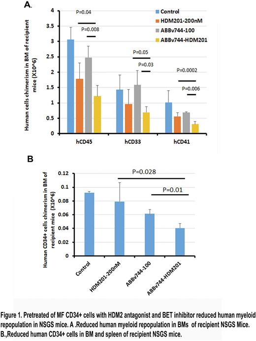
To evaluate the ability of these drugs to eliminate MF HSCs, an immunocompromised NSGS mouse (expressing human IL3, GM-CSF and SCF) xenograft transplantation model was used. MF CD34+ cells were pretreated with vehicle, HDM2a or BETi alone or HDM2a+BETi for 3 days, followed by transplantation into NSGS mice. After 12 weeks, mice were sacrificed and the degree of human cells chimerism in the bone marrows (BM) and spleens of recipient mice evaluated. As compared to vehicle treatment, the mice receiving HDM2a treated grafts had on average 40% less hCD45+ cell chimerism in both organs. BETi treated grafts led to on average a 20% decrease in hCD45+ cells chimerism in the BM but surprisingly not the spleen (Figure 1A). Mice receiving grafts treated with HDM2a+BETi exhibited on average a 60% reduction in hCD45+ cell chimerism in the BM and spleen. Moreover, the grafts treated with HDM2a+BETi generated reduced numbers of cells belonging to multiple hematopoietic lineages (CD34+, CD33+ and CD41+ cells) in both organs of NSGS as compared to mice receiving grafts pretreated with either drug alone indicating that the HDM2a+BETi combination is effective in depleting MF HSCs (Figure 1).
Furthermore, treatment of MF CD34+ cells with WT p53 with an HDM2a or BETi decreased activation of the NFkB pathway, and IL8 and VEGF transcript levels. In addition, treatment of MF CD34+ cells co-cultured with ND mesenchymal stem cells and endothelial cells with the HDM2a+BETi combination led to a 50% reduction in protein levels of IL8, VEGF in the culture supernatants.
These results indicate that combinations of pharmacological agents that disrupt the interplay between HDM2/p53, HIF1α and N-κB pathways could serve as effective therapeutic strategies to treat MF patients.
Disclosures
Hoffman:Novartis: Other: Chair DSMB; Repare: Research Funding; Abbvie: Other: Chair DSMB, Research Funding; Silence Therapeutics: Consultancy; Protagonist Therapeutics: Consultancy; Turning Point: Research Funding; Scholar Rock: Research Funding; Ionis: Consultancy; Novartis: Research Funding.
Author notes
Asterisk with author names denotes non-ASH members.


This feature is available to Subscribers Only
Sign In or Create an Account Close Modal